Dental erosion: A preventive clinical approach
November, 2021.
min read

Dental erosion involves the loss of minerals from tooth structure, caused by acids of non-bacterial origin. This distinguishes erosion from dental caries, which also results in demineralization but that is mediated by the production of acid by cariogenic bacteria.
Erosion occurs when the teeth are in repeated, sustained contact with intrinsic or extrinsic acids.
Intrinsic acids originate in the body. This is most commonly associated with conditions where gastric acid is regurgitated into the oral cavity, like gastroesophageal reflux disease (GERD) or bulimia nervosa. Extrinsic acids originate from outside the body, the most common being dietary acids.
Both intrinsic and extrinsic acids lower the pH of the oral cavity. When the pH level drops below the critical threshold of 5.5, demineralization and softening of the dental enamel will begin to occur.
Saliva has a buffering effect on the oral cavity and offers a degree of protection against enamel erosion. However, this protection can be compromised if salivary flow or buffering capacity is impaired, or if the severity, frequency or duration of exposure to acids exceeds the capacity of saliva.
The tissue softening caused by dental erosion leaves the teeth vulnerable to wear. Attrition may happen during chewing and grinding, and abrasive wear may occur when brushing or cleaning the teeth. The chemical loss caused by the acids together with the activity of attrition/abrasion results in 'erosive tooth wear'.
Accurate diagnosis of dental erosion involves a combination of a visual examination, medical history and a thorough risk assessment.
The Basic Erosive Wear Examination (BEWE) is a helpful tool for identifying and classifying dental erosion. It divides the mouth into six areas, or sextants, and assesses the presence of erosive lesions on every tooth surface. Each sextant is then given a score based on its worst-affected tooth.
0. No erosive tooth wear. The teeth are healthy and the perikymata (faint grooves and ridges) can be seen.
1. Initial loss of surface texture. The perikymata is partially lost. There may be areas of shiny, glossy or dull enamel, and “dimples” or broadened fissures on occlusive surfaces.
2. Distinct defect with hard tissue loss of <50% of the surface area. The surface of the teeth may appear pearly or translucent as erosion advances. There may be areas of yellowish discoloration where the dentin is visible through thinned enamel. You might also see rounded cusps and grooves on incisal edges, or a distinctive rim of enamel at the gingival line.
3. Hard tissue loss of at least 50% of the surface area. You will now see large, distinct areas of tissue loss. The pits and fissures of occlusal surfaces may appear to be smooth and attrition may be visible on the incisors. The patient may also be experiencing dentin hypersensitivity.
During a risk assessment, you should attempt to gain a detailed picture of the patient’s dietary habits, lifestyle, behaviors, and possible medical risk factors.
Examples include:
How often does the patient drink alcohol, sports drinks, soda or fruit juices?
How often does the patient eat acidic foods such as citrus fruits, vinegar or tomatoes?
Does the patient sip drinks slowly, or hold or “swish” drinks in their mouth?
Does the patient drink acidic drinks throughout the day or only with meals?
Does the patient smoke, chew tobacco or use recreational drugs?
Does the patient suffer from conditions such as GERD, bulimia nervosa, alcoholism, obesity, or hyperemesis gravidarum (severe vomiting during pregnancy).
Is the patient taking any medications or undergoing treatment that can cause xerostomia?
Does the patient brush twice a day with a fluoride toothpaste?
Does the patient have any mobility or dexterity issues that may impede proper oral hygiene?
How old is the patient? Is the level of tissue loss commensurate with expected natural loss for their age?
The loss of dental hard tissues associated with dental erosion is irreversible and can have a significant aesthetic and functional impact, so the prevention of dental erosion must always be top of mind. By conducting a visual screening and risk assessment for each patient — not just those who are symptomatic — you can identify at-risk patients and engage the patient in preventing this condition.
1. Educate the patient
Many patients are unaware that their lifestyle and dietary habits can cause dental erosion. For example, your patient might be drinking citrus acidic drinks throughout the day under the impression that this is a “healthy option,” unaware that it could actually be damaging teeth. Prevention and management starts with making your patients aware of dental erosion, the potential long-term consequences, and how their daily habits may contribute to it. Of course, it’s important to provide this education in a compassionate, judgement-free manner to encourage patients to take your advice onboard.
2. Address risk factors
Dietary counselling is one of the most effective tools for preventing dental erosion, but you should also help and encourage your patients to address other risk factors. This may involve:
Treating confounding dental conditions like xerostomia or bruxism.
Referring them to a physician to address contributing medical problems.
Signposting support services for quitting smoking or drug use.
3. Offer effective solutions
There are a number of products, both over-the-counter and prescription, that can support prevention and protection against further dental erosion. Recommend a fluoride toothpaste that supports enamel remineralization and strengthening, like Colgate Enamel Health Toothpaste or the prescription-only Colgate PreviDent 5000 Enamel Protect Toothpaste which protects against acid wear.
Higher-risk patients may benefit from an in-office fluoride varnish treatment like Colgate PreviDent Varnish (or Colgate Duraphat Varnish) with 22,600 ppm fluoride..
4. Provide further resources
Finally, you may wish to offer your patients take-home resources to reinforce your recommendations and advice, or direct them to patient-friendly resources like Mouth Healthy or Colgate.
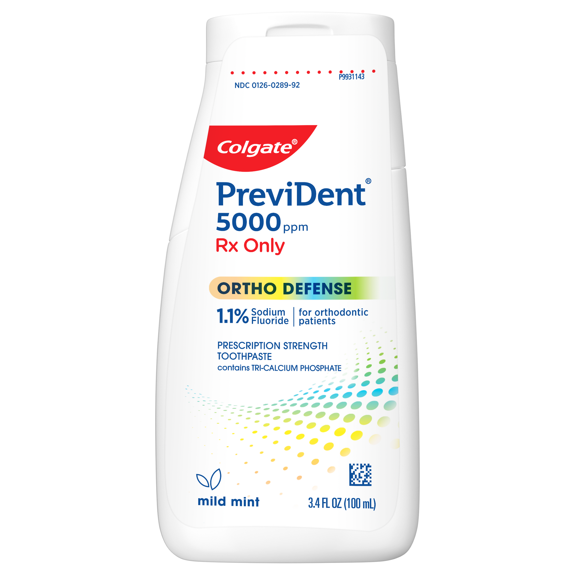
PreviDent® Ortho Defense Prescription Strength Toothpaste helps prevent white spots before they start and keeps your orthodontic patients in optimal oral health. Contact your your dentist today!
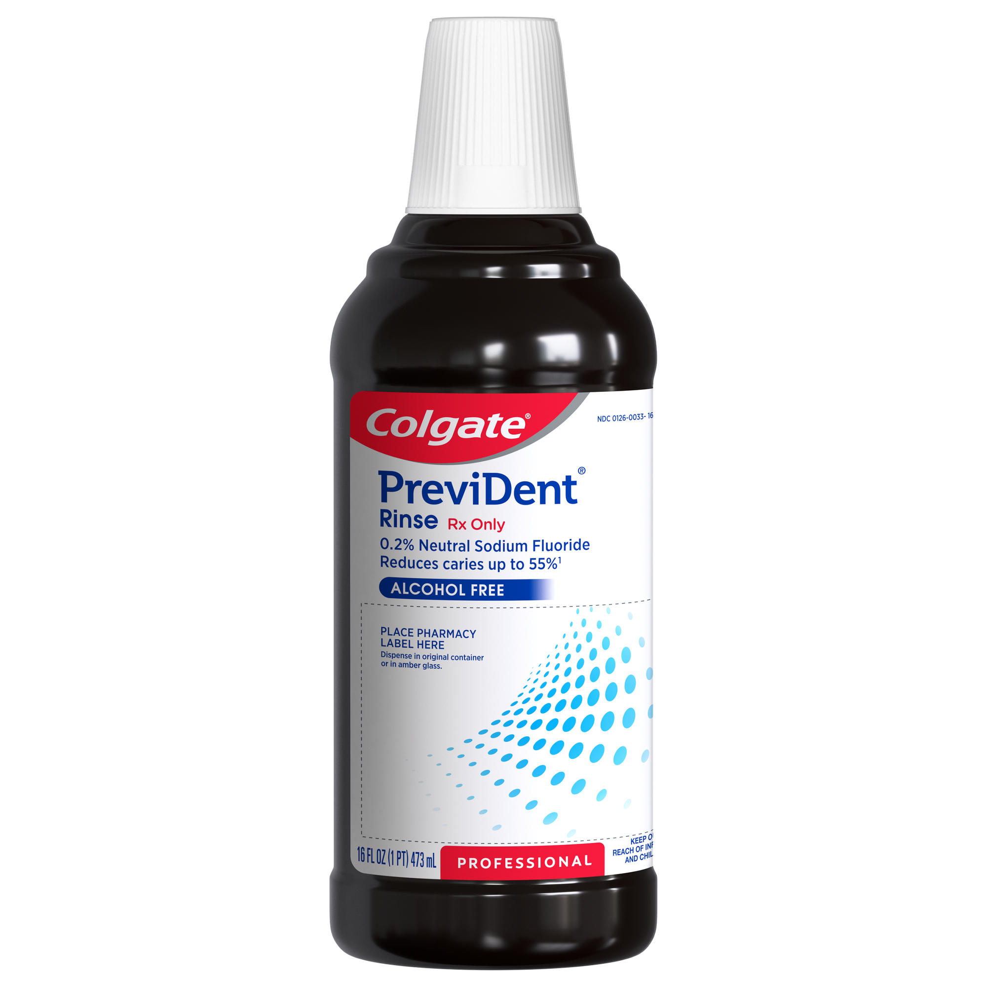
Colgate® PreviDent® Oral Rinse is prescription-strength, with extra fluoride to help decrease cavities for people who have trouble brushing.
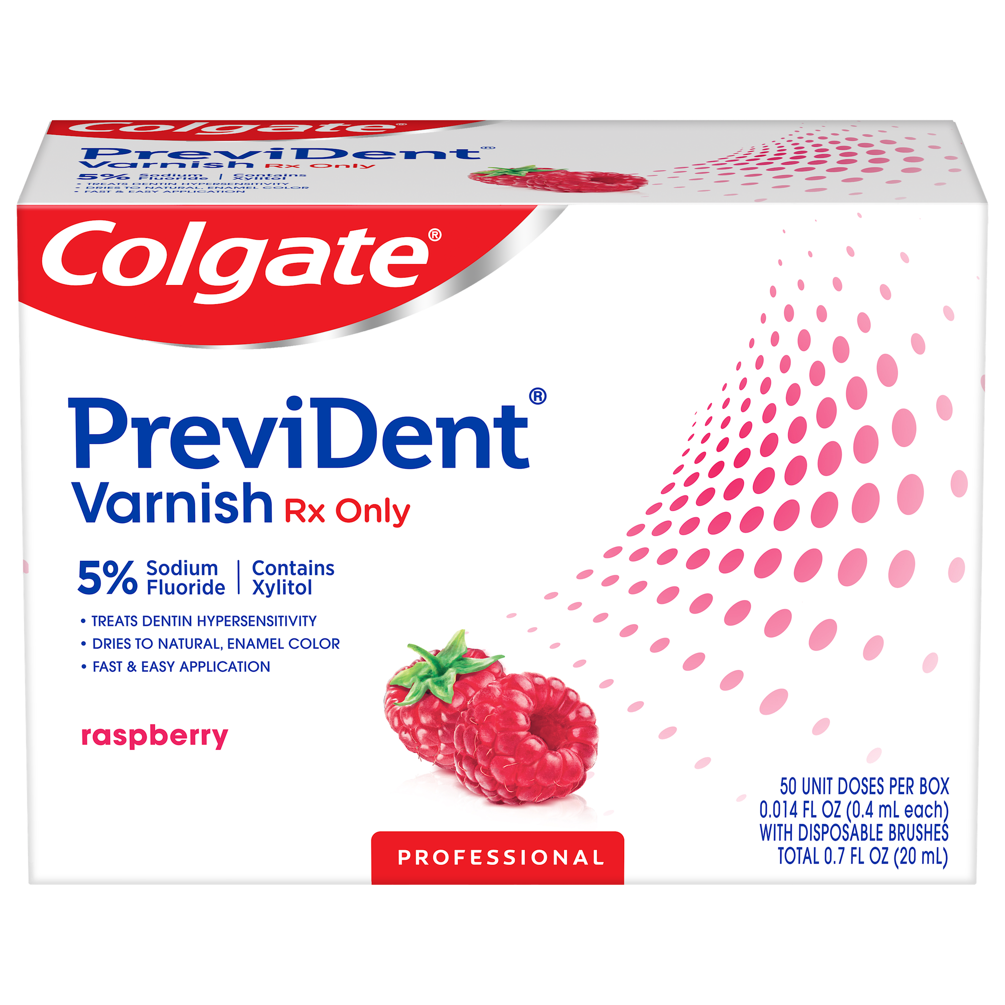
Colgate® PreviDent® Varnish is an in-office treatment for sensitive teeth. Contact your dentist today!

As a dental hygienist, your patients look to you for guidance about their dry mouth symptoms and for suggestions on how to get relief from dry mouth.

Deciding when to use the code D4910 can often be confusing. Patients don't always understand the necessity of this treatment and only want what their insurance covers. As a provider, you want to use the code that matches the treatment provided. Read on for clarification on this topic.

Dietary choices are one of the primary causes of dental erosion. Here’s how to start the conversation with your patient to understand and manage their dietary erosion risk factors.
Get resources, products and helpful information to give your patients a healthier future.
Get resources, products and helpful information to give your patients a healthier future.